
Optic Nerve
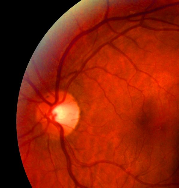 The optic nerve extends all the way from the brain into the skull and the eye. It is also called the nerve of vision. The optic nerve has its major portion enclosed within a bony and rigid tunnel in the skull. Compression or swelling at this location may result in blindness or loss of vision.
Endoscopic optic nerve decompression helps in relieving pressure on the nerve and stabilizing or improving vision. This minimally invasive procedure involves removal of a small portion of the bony optic canal. It is carried out using small rigid telescopes or endoscopes, which allows the surgeon to go through the sinuses and nose for performing this delicate surgery.
Khan Eyelid and Facial Aesthetics, led by oculoplastic & reconstructive surgeon Dr. Tanya Khan, provides safe and proven eye care procedures to patients in Plano, Dallas, Texas, and surrounding locations.
Candidates for Optic Nerve Decompression Surgery
Optic nerve decompression is used for treating conditions that may cause or threaten progressive vision loss because of significant pressure on the optic nerve along the optic canal. The procedure can be used for treating optic nerve pressure because of:
- Fibro-osseous lesions or overgrowth of bone that narrows the optic canal
- Growth or tumors within the eye which exerts direct pressure on the nerve
- Trauma or head injury
Results to Expect
This is a surgical procedure which is performed using general anesthesia. The surgeon will go through the nose to perform an endoscopic sinus surgery. Sinuses open right next to the optic nerve besides the eye. A portion of the optic canal will be removed once this is done.
This creates more space within the bony canal and decompresses the optic nerve. It also reduces pressure on the nerve. Generally, nose packing is not required after the procedure. However, patients may be kept in the hospital overnight for close observation.
Understanding Optical Nerve Sheath Fenestration
Orbital surgeons have preferred approaching the lateral and superior orbit through the anterior superior eyelid crease for removing intraorbital lesions. However, in recent years the anterior superior eyelid crease technique has gained more popularity in neurosurgery. This is because the surgery provides excellent exposure to the anterior skull base.
Excellent cosmetic outcome is a great advantage of the approach. This is because the incision can be easily hidden within the superior eyelid crease. The medial intraconal space can be accessed easily by making an incision in the superomedial eyelid crease for performing an ONSF (Optical Nerve Sheath Fenestration).
The medial horn of the levatoraponeurosis is pushed in place laterally after opening the orbital septum. A plane is created with blunt dissection between the superior oblique tendon and the medial rectus muscle for accessing the posterior orbit.
The optic nerve is then brought into view with further posterior dissection. A rectangular window or a slit is created inside the optic nerve sheath. Decreased traction on the globe, decreased bleeding, decreased operating time, and absence of extraocular muscle dissection is achieved through superiomedial eyelid approach.
Board certified ophthalmologist Dr. Tanya Khan receives patients from Plano, Dallas, Texas, and nearby areas for advanced and innovative eye care treatments.
Contact Khan Eyelid and Facial Aesthetics and Oculoplastic & Reconstructive Surgeon Dr. Tanya Khan Today to Schedule an Appointment
For more information about procedures and treatments at Khan Eyelid and Facial Aesthetics by Ophthalmic surgeon Dr. Tanya Khan. Click here to contact us.
Taking patients from in and around Dallas, Plano, Fort Worth, Grapevine, Garland, Mesquite, Carrollton, Irving, Frisco, Texas and more.
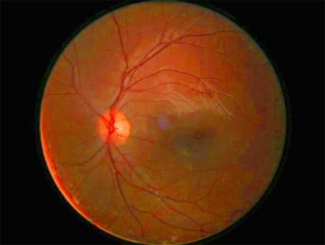 The optic nerve is also called Cranial Nerve II (CN II) or the second Cranial Nerve. It is located at the back of the eye and is responsible for transmitting visual information. The eyes work like the windows to the world with the brain processing the visual information transferred. This is made possible by electric impulses.


Schedule a Consultation: 972-EYE-LIDS (393-5437)