
Contact Us | Location

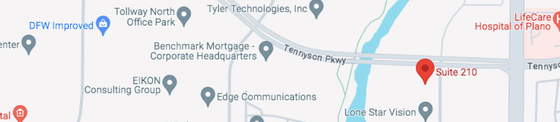
    Dallas
    Khan Eyelid & Facial Aesthetics
    5060 Tennyson Parkway
    Suite 210
    Plano, TX 75024

    Tear Duct Obstruction: Causes, Symptoms, and Treatment Options in Dallas

    Tear Duct Obstruction: Causes, Symptoms, and Treatment Options in Dallas

    Excessive tearing, also called epiphora, is one of the most common complaints that brings patients to an oculoplastic surgeon. While many people assume that tearing simply means their eyes are producing too many tears, the more common cause is a problem with tear drainage, specifically, a blocked or narrowed tear duct. Dr. Tanya Khan, a board-certified ophthalmologist and fellowship-trained oculoplastic surgeon in Dallas and Plano, specializes in the evaluation and treatment of lacrimal (tear drainage) disorders.

    How the Tear Drainage System Works

    Tears are produced by the lacrimal gland and spread across the surface of the eye with each blink. They drain through tiny openings called puncta in the inner corner of each eyelid, then travel through small canals (canaliculi) into the lacrimal sac, and then down through the nasolacrimal duct into the nose. This is why your nose runs when you cry, tears are draining into the nasal cavity. When any part of this drainage pathway is obstructed, tears cannot drain properly and overflow down the cheek.

    Causes of Tear Duct Obstruction

    Age-Related Narrowing

    The most common cause in adults. The tear drainage structures can gradually narrow and eventually obstruct with age. This typically affects women more than men and becomes more common after age 60.

    Nasal or Sinus Disease

    Chronic sinusitis, nasal polyps, or prior nasal surgery can affect the nasolacrimal duct, which passes through bone adjacent to the nasal cavity.

    Infection (Dacryocystitis)

    Acute or chronic infection of the lacrimal sac can occur when tear drainage is obstructed and bacteria proliferate in the stagnant tear pool. Acute dacryocystitis presents with redness, swelling, and pain at the inner corner of the eye and may require antibiotic treatment or surgical drainage.

    Congenital Obstruction

    Nasolacrimal duct obstruction is one of the most common conditions affecting newborns, approximately six percent of infants are born with a membrane blocking the lower end of the nasolacrimal duct. The majority resolve spontaneously within the first year with lacrimal massage.

    Symptoms of Tear Duct Obstruction

    • Persistent tearing or watery eyes
    • Discharge or crusting at the inner corner of the eye
    • Recurrent episodes of redness and swelling at the inner corner of the eye
    • Mucoid or purulent discharge that can be expressed from the tear duct when pressure is applied

    Treatment Options

    DCR (Dacryocystorhinostomy)

    The most common and effective surgical treatment for complete nasolacrimal duct obstruction. DCR creates a new drainage pathway directly from the lacrimal sac into the nasal cavity, bypassing the blocked nasolacrimal duct. It can be performed externally (through a small incision on the side of the nose) or endoscopically (through the nose without external incision). Success rates exceed 90 percent.

    Probing and Irrigation

    For partial obstructions or in infants and children, probing of the tear duct with dilation may restore adequate drainage. Silicone tubes are sometimes placed to maintain the drainage pathway during healing.

    Dr. Khan provides lacrimal system evaluation and DCR surgery to patients in Dallas, Plano, Fort Worth, and throughout North Texas. To schedule a tear duct evaluation, contact Khan Eyelid Surgery today.

    For more information about procedures and treatments at Khan Eyelid and Facial Aesthetics by Ophthalmic surgeon Dr. Tanya Khan. Click here to contact us.

    Taking patients from in and around Dallas, Plano, Fort Worth, Grapevine, Garland, Mesquite, Carrollton, Irving, Frisco, Texas and more.

    Ptosis Repair Surgery: What Dallas Patients Should Know About Droopy Eyelid Correction

    Ptosis, the medical term for drooping of the upper eyelid, is a condition that affects patients across the age spectrum. It can cause a tired or aged appearance, impair peripheral or even central vision in severe cases, and significantly affect quality of life. Dr. Tanya Khan, a board-certified ophthalmologist and fellowship-trained oculoplastic surgeon serving Dallas, Plano, and Fort Worth, specializes in ptosis evaluation and surgical repair.

    What Causes Ptosis?

    Aponeurotic Ptosis

    The most common type in adults. Caused by stretching or detachment of the levator aponeurosis, the tendon connecting the levator muscle to the eyelid. This is age-related, progressive, and may be accelerated by contact lens wear or eye rubbing. The levator muscle itself is intact; it is the attachment that has weakened.

    Myogenic Ptosis

    Caused by a disorder of the levator muscle itself, such as chronic progressive external ophthalmoplegia or myasthenia gravis. Identifying and treating the underlying neuromuscular condition is an important component of management.

    Neurogenic Ptosis

    Caused by a problem with the nerve supply to the levator muscle. Horner’s syndrome and third nerve palsy are the most common causes. Ptosis of sudden onset, particularly if associated with double vision, headache, or other neurological symptoms, requires urgent evaluation.

    Congenital Ptosis

    Present from birth, usually due to maldevelopment of the levator muscle. Congenital ptosis requires prompt evaluation and treatment in children to prevent amblyopia (lazy eye) from developing due to visual axis obstruction.

    Ptosis vs. Excess Eyelid Skin (Dermatochalasis)

    This is one of the most important distinctions in oculoplastic surgery. Ptosis is drooping caused by a problem with the muscle or its attachment, the eyelid margin itself is low. Dermatochalasis is excess upper eyelid skin that overhangs the lashes, but the eyelid margin is in a normal position. The treatment is different: ptosis repair elevates the eyelid margin; blepharoplasty removes excess skin. Some patients have both and benefit from both procedures.

    Ptosis Repair: Surgical Options

    Levator Advancement

    The most common surgical approach for aponeurotic ptosis. The surgeon tightens or reattaches the levator aponeurosis to the eyelid through an incision in the upper eyelid crease. This is typically performed under local anesthesia so that the patient can open their eyes during surgery, allowing precise height and contour adjustment.

    Mullerectomy (Posterior Approach)

    An internal approach that tightens Muller’s muscle from behind the eyelid. Performed in patients with good levator function who respond to phenylephrine eye drops.

    Frontalis Suspension

    For patients with very poor levator function, including many cases of congenital ptosis, the eyelid is suspended from the frontalis brow muscle using a sling.

    Insurance Coverage

    Ptosis repair is a functional procedure when the drooping eyelid is causing documented visual field obstruction. In these cases, health insurance, including Medicare, typically covers the surgery. Dr. Khan’s team assists patients with the documentation and prior authorization process.

    Dr. Khan provides ptosis repair and eyelid surgery to patients in Dallas, Plano, Fort Worth, Austin, and throughout North Texas. To schedule an evaluation, contact Khan Eyelid Surgery today.

    For more information about procedures and treatments at Khan Eyelid and Facial Aesthetics by Ophthalmic surgeon Dr. Tanya Khan. Click here to contact us.

    Taking patients from in and around Dallas, Plano, Fort Worth, Grapevine, Garland, Mesquite, Carrollton, Irving, Frisco, Texas and more.

    ABO AAO RealSelf